Why Lyophilisation Matters for Peptide Stability
Research-grade peptides are supplied in lyophilised form (a freeze-dried powder produced by removing water from the peptide solution under vacuum at low temperature). The process is not merely a storage convenience; it is an active preservation strategy that exploits the relationship between water activity and molecular degradation rates. In aqueous solution, peptides are subject to hydrolysis of peptide bonds, oxidation of susceptible residues, and aggregation driven by hydrophobic interactions. Remove the water and all three processes slow to negligible rates at room temperature.
How the freeze-drying process preserves peptide structure explains the physical chemistry in more detail, but the practical implication is clear: the lyophilised cake in a research vial is the stable form, and the reconstituted solution is inherently less stable. Once water is reintroduced, the degradation clock starts. Everything that follows in this protocol is designed to slow that clock as much as possible.
The appearance of lyophilised peptide, a white to off-white powder or compressed cake, gives no reliable indication of purity or activity. Quality determination requires analytical data from the supplier. What the visual inspection can tell you is whether the vial has been compromised: a wet or clumped cake suggests moisture ingress, which would have begun the degradation process before reconstitution.
Bacteriostatic Water vs Sterile Water — When Each Is Correct
The choice of diluent is the single most consequential decision in reconstitution. Sterile water for injection (SWFI) is purified water with no added preservatives. It is appropriate for single-use reconstitutions where the entire contents of the vial will be used immediately. Because it contains no antimicrobial agent, multi-puncture use is microbiologically unsafe: each needle insertion risks introducing bacterial contamination that will not be inhibited.
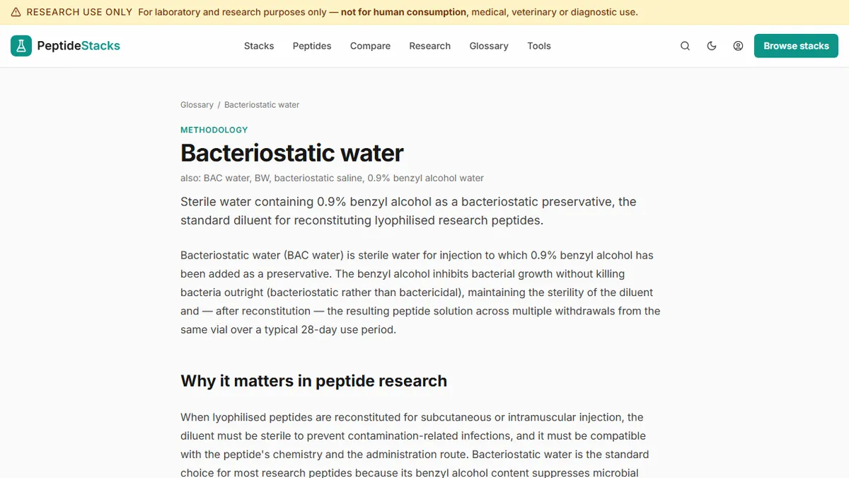
Bacteriostatic water for injection (BWFI) contains 0.9 percent benzyl alcohol as a preservative. This antimicrobial agent prevents the growth of most common bacterial contaminants and makes the reconstituted solution safe for multiple punctures over its 28- to 30-day usable window. For research protocols involving repeated draws from the same vial, which is the standard pattern when a compound is dosed twice daily over several weeks, bacteriostatic water is the correct choice.
Why bacteriostatic water is the standard diluent for research peptides covers the antimicrobial chemistry in full. The practical point is this: using sterile water for a multi-dose vial and then storing it in the refrigerator for two weeks is an unacceptable microbiological risk in any research context.
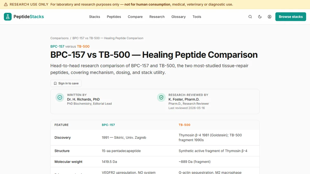
One exception worth noting: some peptides are sensitive to benzyl alcohol at high concentrations. This is documented for certain cytokine-adjacent compounds but is not a significant concern for the most commonly researched peptides including BPC-157, TB-500, and the GLP-1 agonist analogues. If working with a less common compound, check the specific stability data.
Step-by-Step Reconstitution: Alcohol Swab, Slow Wall Injection, No Vortexing
Before beginning, assemble the necessary materials: the lyophilised peptide vial, bacteriostatic water, a sterile syringe for drawing the diluent, a sterile syringe or needle for the reconstituted solution, and alcohol swabs. Work on a clean surface; a laminar flow hood is the ideal environment for research applications, but at minimum the workspace should be free of airflow and contamination sources.
Step one: wipe the rubber stopper of both the bacteriostatic water vial and the peptide vial with fresh alcohol swabs and allow them to dry for at least ten seconds. Alcohol requires contact time to be effective; wiping and immediately puncturing defeats the purpose.
Step two: draw the required volume of bacteriostatic water into the syringe. For most peptides, 1 mL or 2 mL is the standard reconstitution volume, and the choice of volume determines the resulting concentration. Calculate the target concentration before drawing the diluent.
Step three: insert the needle into the peptide vial and direct the stream of bacteriostatic water against the inside wall of the vial rather than directly onto the lyophilised cake. This wall-injection technique is critical. Forceful direct contact with the powder can disrupt the secondary and tertiary structure of the peptide chains, potentially reducing biological activity. The water should run down the wall and dissolve the cake gently from the base up.
Step four: once the diluent has been introduced, allow the vial to sit undisturbed for two to three minutes. Gentle swirling, a slow rotation of the vial between the fingers, is acceptable. Vortexing, shaking, or any vigorous mechanical agitation is not. The shear forces generated by vortexing can cleave peptide bonds and aggregate the compound. The solution should become clear and colourless or faintly yellow; cloudiness indicates aggregation or incomplete dissolution.
Calculating Concentration and Filling the Syringe Accurately
The concentration of the reconstituted solution depends on two variables: the mass of peptide in the vial and the volume of diluent added. If a vial contains 5 mg of peptide and 1 mL of bacteriostatic water is added, the resulting concentration is 5 mg per mL, or 5,000 mcg per mL. A 500 mcg dose from this solution requires drawing 0.1 mL.
When using a U-100 insulin syringe (the standard syringe type for subcutaneous peptide administration in research protocols), the markings represent units calibrated for U-100 insulin. Each unit on the syringe equals 0.01 mL. A 0.1 mL draw corresponds to the 10 unit mark. Researchers unfamiliar with U-100 syringes sometimes confuse units with microlitres or microgram doses; the safest approach is always to calculate the required volume in millilitres first, then identify the corresponding syringe marking.
Working examples: BPC-157 reconstituted at 1 mg per mL yields a 500 mcg dose at 0.5 mL, which is the 50 unit mark on a U-100 syringe. TB-500 reconstituted at 2.5 mg per mL yields a 2.5 mg dose at 1 mL.
Storage Conditions — 2-8 Celsius, Light Protection, 30-Day Limits
Reconstituted peptide solutions should be refrigerated at 2 to 8 degrees Celsius immediately after preparation and kept there for the duration of use. This temperature range slows but does not halt the hydrolytic and oxidative degradation processes that resume upon reconstitution. Most peptides reconstituted in bacteriostatic water maintain acceptable stability for 28 to 30 days under refrigeration; longer storage is not supported by available stability data for most compounds.
Light protection is a secondary but real concern for several peptides, including GHK-Cu. The copper coordination in GHK-Cu is susceptible to photochemical reactions that can alter the oxidation state of the cupric ion and degrade the peptide-metal complex. Amber vials or storage in a dark drawer addresses this. For compounds without specific photosensitivity documented, the refrigerator's internal darkness provides adequate light protection incidentally.
Freeze-thaw cycling degrades most peptide solutions progressively: ice crystal formation can fragment peptide chains, and the concentration-dilution dynamics of freezing and thawing shift the chemical equilibrium in ways that promote aggregation. If a compound will not be used within the 30-day window, the preferred approach is to aliquot the reconstituted solution into single-dose vials before freezing rather than freezing and re-thawing the main vial repeatedly.
Signs a Reconstituted Peptide May Have Degraded
Several visual and physical indicators suggest that a reconstituted peptide solution should be discarded rather than used. Cloudiness or visible particulate matter indicates aggregation: the peptide chains have clumped into insoluble assemblies that will not reconstitute by swirling. Colour change beyond the faint yellow that some compounds naturally exhibit at reconstitution suggests oxidative degradation. Unusual odour is a non-specific but meaningful signal that the chemistry of the solution has changed.
Beyond visual inspection, solutions stored beyond the 30-day window or exposed to temperature excursions above 8 degrees for extended periods should be treated as potentially degraded regardless of appearance. Degradation products may not change the visual character of the solution but can have different biological activities — or none at all. In a research context, using degraded material introduces a confounding variable that invalidates the experimental result.